face reconstruction
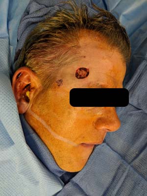
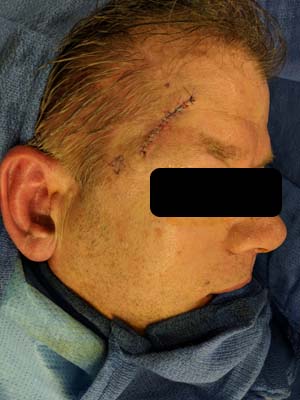
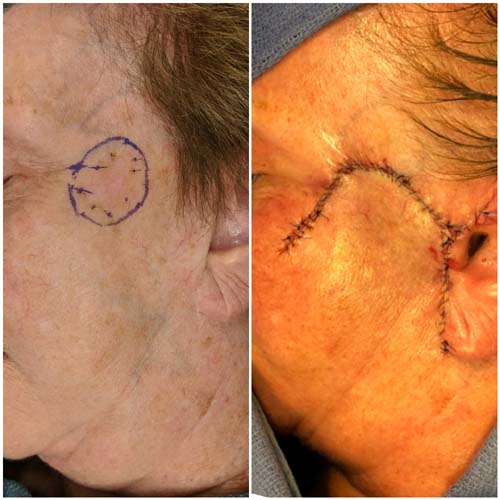
Case 2: The patient is shown immediately after excision of a skin cancer on the temple, and is seen immediately after closure of the skin defect.
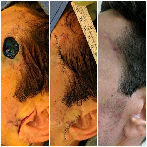
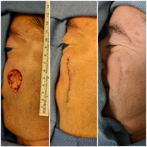
Case 3: The patient is shown immediately after excision of a skin cancer on the temple, and is seen immediately after closure of the skin defect in the operating room, and then 3 weeks after the operation at a clinic follow up visit.
Case 4: The patient is shown immediately before excision of a skin cancer of the cheek, and is seen immediately after closure of the skin defect in the operating room with movement of local skin to fill the defect.
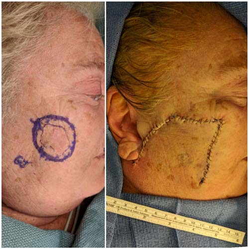
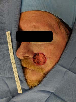
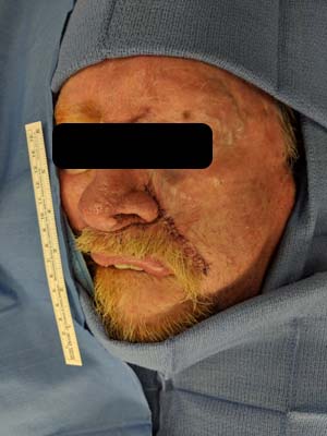
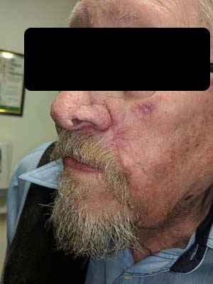
Case 5: The patient is shown immediately before excision of a skin cancer of the face, and is seen immediately after closure of the skin defect in the operating room with movement of local skin to fill the defect.
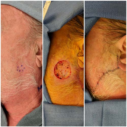
Case 6: The patient is shown immediately after excision of a skin cancer of the cheek, and is seen immediately after closure of the skin defect in the operating room, and then in follow up 2 weeks later.
Case 7: The patient is shown immediately before and after excision of a skin cancer of the the junction between the cheek and the jawline, and is seen immediately after closure of the skin defect in the operating room.
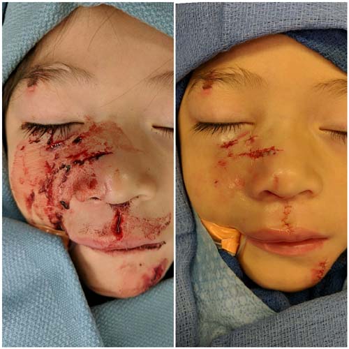
Case 8: A child is shown immediately before and after repair of multiple dog bites to the face in the operating room.
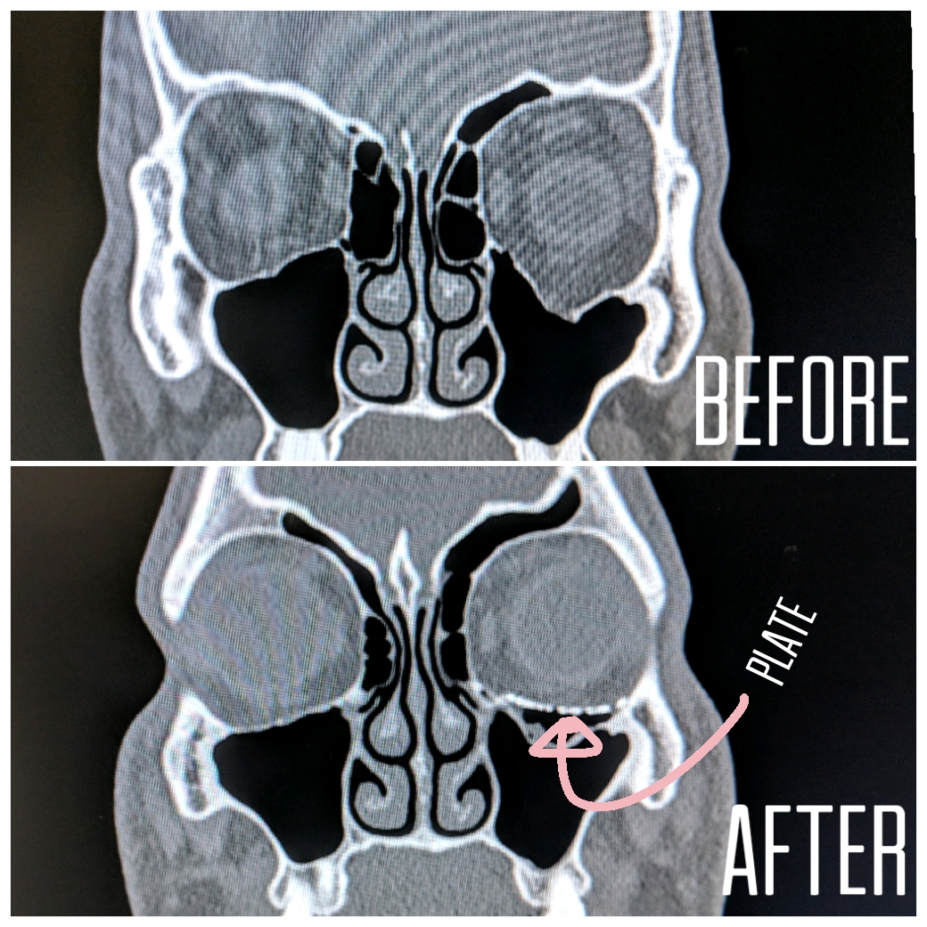
Case 9: A fracture of the orbital floor (base of the eye) is shown on a CT scan before and after repair with a titanum mesh plate.